An experimental PET radiotracer called Ga-68 NK224 can detect programmed death ligand 1 (PD-L1) expression across the entire body in lung cancer patients, providing a more complete picture of disease than traditional biopsies and helping predict which patients will respond to immunotherapy treatment.
PET/CT scans with an experimental radiotracer that targets programmed death ligand 1 (PD-L1) protein on cancer cells can reveal the full burden of disease in patients with non-small cell lung cancer, according to a study published August 2026 in Radiology.
A group from the First Affiliated Hospital of Xiamen University in Xiamen, China, performed gallium-68 (Ga-68) NK224 PET/CT scans in participants with newly diagnosed, recurrent, or metastatic non-small cell lung cancer (NSCLC), with findings suggesting the approach can overcome inherent sampling limitations of single-site biopsies. Ultimately, the modality may aid personalized immunotherapy strategies for patients with multifocal metastatic disease, the authors suggested.
“Ga-68 NK224 PET/CT enabled whole-body assessment of programmed death ligand 1 heterogeneity in participants with non–small cell lung cancer and revealed lesion-level heterogeneity not captured through biopsies,” noted lead authors Liang Zhao, MD, and Hui Zhou, MD, and colleagues. “Ga-68 NK224 uptake was associated with immunotherapy response.”
Currently, PD-L1 immunohistochemical analysis performed on biopsy specimens is the standard method for evaluating patients for immunotherapy. However, increasing evidence indicates that PD-L1 expression exhibits pronounced intratumoral and intertumoral heterogeneity, which can make immunotherapy decision-making based on biopsies challenging, the researchers explained.
Alternatively, molecular imaging can provide whole-body visualization and quantitative assessment of PD-L1 expression, and to that end the group previously developed Ga-68 NK224 and found it safe and effective in a small group of patients. In this study, the researchers tested it in a larger cohort in a prospective study from December 2023 to July 2025.
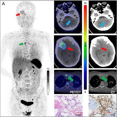
According to the findings, Ga-68 NK224 uptake differed significantly across PD-L1 tumor proportion score (TPS) categories (based on the percentage of positive tumor cells in the biopsy stain) using both PET biopsy-plane (local) and whole-lesion regions of interest (ROIs) (both p < .001). A maximum standard uptake value (SUVmax) cutoff of 5.6 best discriminated high PD-L1 expression, and among 12 participants classified as PD-L1 negative by immunohistochemical analysis, five had at least one lesion exceeding this threshold.
Intratumoral heterogeneity was evident, with greater Ga-68 NK224 uptake variability in whole-lesion ROIs than biopsy-plane ROIs (median SUVmax 1.0 versus 0.4; p < 0.001) and intertumoral heterogeneity within individual participants was substantial (median SUVmax coefficient of variation, 23.6%). Furthermore, among 24 lesions from immunotherapy-treated participants, responders showed greater Ga-68 NK224 uptake than non-responders (median SUVmax, 5.9 vs 1.9), the researchers reported.
“By overcoming the inherent sampling limitations of single-site biopsy, Ga-68 NK224 PET/CT provides a more comprehensive assessment of tumor immune phenotypes,” the authors wrote.
Validating quantitative uptake thresholds and determining whether PD-L1 PET-guided treatment selection improves outcomes will require larger multicenter and multiethnic trials, they concluded.