Optical guided precision biological X-ray irradiator can mark tumor cells by bioluminescence, mark tumor drugs by fluorescent dyes, and locate tumor drugs by X-ray. It can identify the exact location of the tumor, whether the drugs reach the tumor in a targeted way, as well as the drug metabolism. At the same time, it uses X-ray to accurately focus on the tumor location, and implements accurate radiotherapy using X-ray's strong tissue penetration ability, thus providing an ideal treatment plan.
Optical guided precision biological X-ray irradiator provides information about the location, size, shape and other aspects of the tumor by optical means. However, on the basis of not increasing the toxic and side effects of the surrounding normal tissues, to further improve the therapeutic effect is still an urgent problem to be solved in the field of tumor radiotherapy.
X-ray photosensitizers can be used as one of the important methods to solve the above problems by virtue of great potential and excellent advantages in material research. They can be effectively enriched to the tumor site through molecular targeting ligands, and activated in deep tumors through X-ray tissue penetration. Materials with different properties can load a variety of photosensitizers and help X-ray energy transfer to photosensitizers, so that X-ray photosensitizers can be excited to achieve ideal therapeutic effects.
Self-shielding large box body design, the ability to accommodate a wide range of accessories, OptiMAX multi-modal imaging system (including X-ray imaging, bioluminescence and fluorescence)
TouchRAD 15″ super touch screen, user-friendly interface, configuration of uses-permission and data management rights, Windows operating system, automatic generation of Excel data, USB interface to facilitate data export.
● Equipped with a variety of filters, supporting multi-wavelength data acquisition
● Multi-modal optical imaging system: a high-cooling, high-sensitivity EMCCD camera for optical/luminescent presentation, which can capture images at any angle,EMCCD sensor, with a resolution up to 0.2mm, easy to fuse with CT imaging, and its optical aiming accuracy:<1mm.
● X-ray tube: COMET, maximum power 4000W, automatic preheating
● Tube type: cermet, fixed anode, water cooled
● X-ray energy range: 5-225 kV
● Adjustable irradiation distance: 15-90 cm SSD
● Focus size: 7.5 mm (per EN12543)
● Main unit size (width × depth × height): 142cm × 78cm × 193cm
● Irradiation chamber size (width × depth × height): 64cm × 69cm× 106cm
● Weight: 1497 kg
● Power requirements: 230VAC,50A,50/60 Hz
● Cooling pump: water-air
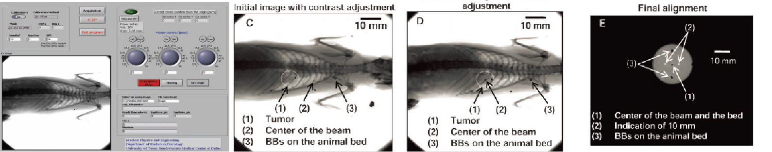
B: Software for image acquisition and positioning. C: Initial positioning image indicating beam center and tumor. D: Verification of the second image. E: Calibration and centering relative to the beam axis.