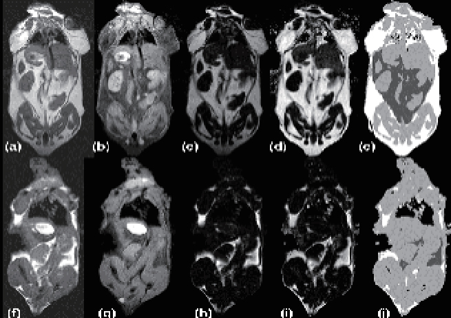
Magnetic resonance imaging (MRI) products can visually show the specific distribution of adipose tissue. By selecting different experimental angles, we can analyze the distribution area of adipose tissue at different locations, whether there is variation in adipose tissue at different sites or in organs, and whether the adipose tissue in the same organic organs bilaterally is symmetrically distributed. The imaging device can help better assess anatomical and disease morphology, physiological and functional parameters, and tissue system study, such as adipose tissue, skeletal joint and soft tissue, and etc.
Features:
1. It can realize magnetic resonance imaging of small animals such as rats, mice, rabbits, small dogs, monkeys, etc., and provide MRI solutions for small animals at preclinical and molecular level.
2. By combining advanced RF coil and gradient technology with ultra-high field magnetism, our system can provide the spatial resolution of living organisms.
3. Extensible to add PET and SPECT modules.
In vivo fat study
In the image, non-visceral fat tissue is white, visceral fat is dark gray, muscles and organs are light gray, and air (lungs/background) is black.